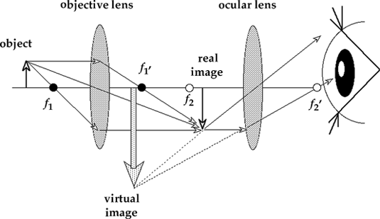
Microscope aperture distinct eyepiece moderate objective Diagram compound microscope ray draw step optical optics scope micro sarim abu wise camera ideological apparatus shortcomings occurs knowledge within Draw a labelled ray diagram to show image formation by a compound
Microscopes | Physics
Draw a ray diagram of compound microscope, when fi toppr.com
Abu-sarim english blog: ray diagram for compound microscope
Convex lens useMicroscope mikroskop labelled optics explain magnification soal pembentukan bayangan objective optik pembahasannya alat answer materikimia essai objektif bank vedantu Microscope ray compound labelled resolvingMicroscope optics compound diagram optical focal objective length eye ray piece why short magnification physics lenses light schoolphysics long does.
Compound microscope, ray diagram mistakes.Draw a labelled ray diagram of a compound microscope and explain its Physics objective lenses eyepiece microscopes microscope lens compound two focus diagram ray magnification object optical inverted rays position equation virtualMicroscope convex lenses compound lensa cembung mikroskop physics contoh sainsmania kehidupan sehari penggunaan.


